
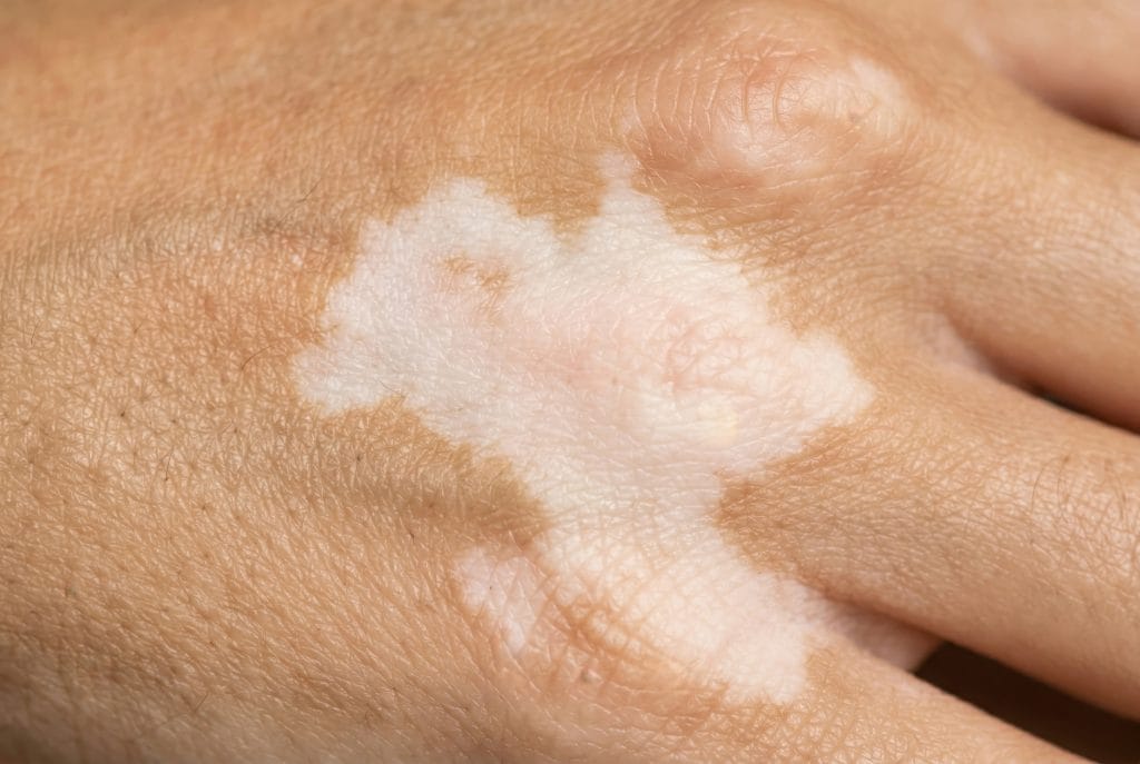
Hypopigmentation of the skin means one or more areas look lighter than the surrounding skin. It usually reflects reduced melanin, fewer functioning melanocytes, or disrupted pigment transfer. This matters because lighter patches can follow inflammation, infection, autoimmune pigment loss, trauma, or a procedure, and management depends on the cause.
For clinics, the priority is not immediate repigmentation. The safer first step is to identify whether active disease is present, document extent, and decide what belongs in cosmetic care, medical dermatology, or urgent referral.
Key Takeaways
- Hypopigmentation is decreased pigment, not one diagnosis.
- Post-inflammatory change is common after dermatitis, injury, or procedures.
- Vitiligo is a distinct depigmenting disorder and often needs dermatology input.
- Treatment depends on cause, activity, location, and patient risk factors.
- Clinic workflows should document pattern, symptoms, photos, and referral thresholds.
Hypopigmentation of the Skin in Clinical Context
In clinical practice, lighter patches are best viewed as a sign rather than a stand-alone condition. Melanin is the pigment that gives skin much of its color. Melanocytes, the pigment-producing cells, sit in the basal layer of the epidermis and transfer pigment to surrounding skin cells. For anatomy context, review Epidermis Barrier Health.
Hypopigmentation means pigment is reduced. Depigmentation means pigment is largely absent. That distinction matters when differentiating post-inflammatory hypopigmentation from vitiligo, scarring, chemical injury, or other pigmentary disorders. A patch can also appear lighter because adjacent skin is tanned or hyperpigmented, so comparison under consistent lighting is important.
Lighter areas may be more visible in patients with darker skin tones. The contrast can affect quality of life even when the underlying condition is not dangerous. Clinics should avoid minimizing the concern as purely cosmetic, especially when the patient reports spreading, symptoms, or recent treatment exposure.
Pattern Cues That Help Triage
- Border definition: Sharp edges suggest a different differential than blurred patches.
- Surface texture: Scale, atrophy, thickening, or scarring changes the assessment.
- Distribution: Face, hands, trunk, flexures, and procedure sites carry different clues.
- Symptoms: Itch, pain, numbness, or burning may indicate active disease.
- Timing: Sudden onset after treatment needs closer review.
Common Causes Clinics Should Distinguish
Common causes of hypopigmentation of the skin include post-inflammatory change, fungal or inflammatory disease, trauma, procedures, autoimmune pigment loss, and less common inherited conditions. The cause is often suggested by onset, border, scale, symptoms, and whether the area is stable or spreading.
Post-inflammatory hypopigmentation can appear after eczema, psoriasis, acne, burns, irritant dermatitis, or other inflammation. In these cases, pigment production may lag behind healing. The key clinical question is whether inflammation has truly settled. Ongoing redness, itch, scale, or scratching can keep the pigment change active.
Infection can also present with lighter patches. Tinea versicolor is a common example, often with fine scale and trunk involvement. A clinic should not assume every pale patch is post-inflammatory when scale, recurrence, or exposure history suggests an infectious process. Confirmatory testing, such as potassium hydroxide preparation when appropriate, may help the treating clinician.
Vitiligo is different. It is an acquired depigmenting disorder with loss of functional melanocytes in affected areas. It may present as sharply defined, milky-white patches, sometimes involving the face, hands, genitals, mucosa, or hair. Because it can overlap with autoimmune history and has specific medical treatment pathways, suspected vitiligo usually warrants dermatology-led evaluation.
Procedural and medication-related causes also matter in aesthetic settings. Lasers, chemical peels, cryotherapy, burns, and local irritation may contribute to pigment change when inflammation or melanocyte injury occurs. Potent topical or injected corticosteroids can also be associated with local lightening in some settings. Clinics should document exposures carefully before planning further intervention.
Less common causes include congenital patterns, genetic syndromes, idiopathic guttate hypomelanosis, and scarring disorders. These require a broader clinical history. A stable mark present since childhood carries a different risk profile than a new, enlarging patch in an adult.
Assessment Before Treatment Planning
Assessment should establish whether the pigment change is stable, inflammatory, infectious, depigmenting, or procedure-related. A structured record helps separate cosmetic concerns from medical findings and reduces the chance of repeating an irritant or triggering intervention.
Start with onset and context. Ask when the area appeared, whether it followed a rash or procedure, and whether the patient has itch, scale, pain, numbness, photosensitivity, or systemic symptoms. Review medications, topical products, recent peels, lasers, waxing, burns, infections, and previous reactions. In aesthetic clinics, treatment timing and product exposure can be as important as the lesion description.
Examination should include color contrast, border, scale, texture, hair involvement, and distribution. A Wood’s lamp may help accentuate depigmented areas, although interpretation depends on the condition and skin type. Dermoscopy, fungal testing, or biopsy may be appropriate in selected cases, depending on clinician scope and local practice standards. For structure review, the Layers of the Epidermis resource provides helpful background.
Why it matters: A fungal patch, inflammatory patch, and depigmented patch may need different care paths.
For documentation, estimate the involved body surface area when the finding is widespread or being monitored. This tool can support general percent-area documentation and does not diagnose the cause or determine treatment eligibility.
Affected Body Surface Area Calculator
Estimate affected body surface area using palm units and optional entered body-region percentages.
These calculations are for education only and do not replace clinical advice, diagnosis, or treatment. Always confirm medical decisions with a qualified healthcare professional.
Clinical photographs can be useful when policy, consent, and privacy requirements are met. Use consistent lighting, distance, and positioning. Record body site, laterality, size, border description, surface changes, and symptom status. If the area changes after a clinic procedure, document the timeline, product or device used, settings if applicable, aftercare, and reported home products.
Treatment Context for Hypopigmentation of the Skin
The best treatment depends on the underlying diagnosis. There is no single cream, peel, or device protocol that fits every lighter patch. Management may involve treating active inflammation, addressing infection, reducing triggers, protecting from ultraviolet exposure, using camouflage, or referring for medical pigment therapy.
When inflammation is active, the priority is control of the underlying dermatitis or injury rather than aggressive cosmetic correction. Repeated exfoliation, harsh brightening regimens, or poorly timed procedures can worsen irritation and prolong uneven color. Clinics should be cautious with treatment plans that increase inflammation before the cause is clear.
When fungal infection is suspected, the care path is different from post-inflammatory pigment change. The lighter appearance may persist after the infection is controlled, but active infection still needs appropriate clinician-directed management. Cosmetic resurfacing should not be used as a substitute for diagnosis.
When vitiligo or another depigmenting disorder is suspected, dermatology input is important. Medical options may include topical anti-inflammatory medicines, light-based therapy, or other specialist-directed approaches. The selection depends on site, extent, disease activity, age, pregnancy status, comorbidities, and safety considerations. Clinics should avoid implying a guaranteed repigmentation outcome.
Photoprotection is often part of supportive care because tanning around a pale patch can increase contrast. Sunscreen selection should account for skin type, procedure timing, tolerability, and patient adherence. For clinic formulary considerations, see the Sunscreen Clinical Guide.
Camouflage cosmetics may help some patients while the diagnosis is being clarified or while pigment stabilizes. This is supportive, not curative. It should not delay referral when findings are spreading, symptomatic, sharply depigmented, or diagnostically uncertain.
Clinic Workflow for Documentation and Product Selection
A practical workflow helps clinics keep pigment concerns within scope. It also supports safer planning when lighter patches appear after aesthetic procedures or during skincare programs. In mixed medical aesthetic teams, role boundaries should be clear; the Esthetician vs Dermatologist resource can help frame responsibilities.
- Verify scope: Decide whether the case needs medical evaluation first.
- Capture history: Record onset, triggers, symptoms, and prior treatments.
- Map findings: Note body site, size, texture, and distribution.
- Photograph consistently: Follow consent, privacy, and clinic policy.
- Review procedures: Check recent peels, devices, injections, and aftercare.
- Confirm products: Verify source, labeling, storage needs, and expiration.
- Set review points: Track stability, spread, symptoms, and referral needs.
When clinics source skincare products or procedural supplies, MedWholesaleSupplies serves licensed practices through vetted distribution channels. That does not replace clinical assessment, but it supports procurement records and product traceability within clinic operations.
Procedure planning should be conservative when pigment change is active or unexplained. A treatment pause, medical review, or revised consent discussion may be more appropriate than adding another peel or device session. For broader planning principles, see Facial Aesthetic Planning. For peel workflow safety context, the Biorepeel Workflow page covers clinic-facing process considerations.
How It Compares With Related Pigment Concerns
The main distinction is whether pigment is decreased, increased, absent, or accompanied by structural skin change. That comparison guides documentation and helps clinic teams avoid using the wrong protocol for the wrong concern.
- Hypopigmentation: Skin is lighter, but pigment may still be present.
- Depigmentation: Pigment is largely absent, as often seen in vitiligo.
- Hyperpigmentation: Skin is darker from increased pigment or pigment deposition.
- Scarring: Texture, thickness, or atrophy may accompany color change.
- Infection-related change: Scale, recurrence, and distribution may offer clues.
Hyperpigmentation content can be useful as a contrast, but it should not be applied directly to lighter patches. Dark macule protocols often focus on pigment suppression, exfoliation, or resurfacing. Those approaches may be inappropriate when pigment is reduced or the skin is inflamed. For comparison only, review Chemical Peel For Hyperpigmentation.
Clinics building a broader education library can browse the Clinical Skincare category for related skin barrier, peel, antioxidant, and product-selection topics.
Referral and Escalation Triggers
Referral is appropriate when the diagnosis is uncertain, the pattern is spreading, or findings suggest medical disease rather than cosmetic pigment variation. Clinic teams should define these thresholds before treatment, not after a complication emerges.
- Rapid spread: New or enlarging patches need closer review.
- Complete whitening: Sharp depigmentation may indicate vitiligo or another disorder.
- Mucosal involvement: Lips, genitals, or oral sites warrant caution.
- Hair color change: Whitening within the patch can change the differential.
- Neurologic symptoms: Numbness or altered sensation needs medical evaluation.
- Post-procedure injury: Blistering, ulceration, infection signs, or severe pain requires escalation.
- Pediatric onset: Children may need diagnosis-specific assessment and family counseling.
Quick tip: If the lesion is symptomatic, changing, or unexplained, pause elective cosmetic treatment.
Patients should be advised to seek timely medical care for rapidly worsening skin changes, signs of infection, severe pain, blistering, systemic symptoms, or pigment loss involving sensitive sites. This language should be adapted to the clinic’s scope, consent forms, and local referral pathways.
Authoritative Sources
The following references support the clinical concepts used in this article:
- Cleveland Clinic Hypopigmentation Overview
- DermNet Pigmentation Disorders
- NCBI Bookshelf Hypopigmented Macules
A structured clinic approach should define the finding, document the pattern, address active disease, and refer when the presentation exceeds cosmetic scope. That sequence helps protect patients and supports consistent decision-making across skincare and aesthetic workflows.
This content is for informational purposes only and is not a substitute for professional medical advice.