

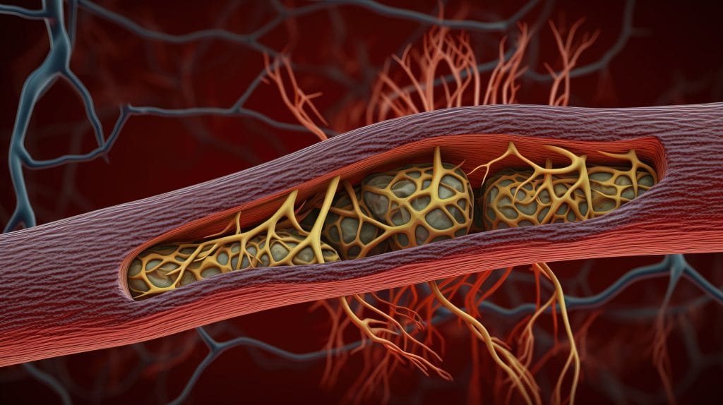
A vascular occlusion is a partial or complete blockage of blood flow in a vessel. In clinic-facing injection practice, it matters because reduced perfusion can injure skin, eyes, nerves, or other tissues if warning signs are missed. The cause may be a clot, an embolus, external pressure, or injected material entering or pressing on a vessel. Licensed clinicians need a clear recognition and escalation plan, not a search-snippet definition. This article focuses on signs, risk context, and operational safeguards relevant to injectable procedures and procurement workflows.
Key Takeaways
- A vessel blockage can affect arteries or veins; consequences depend on location, duration, and tissue involved.
- Suspected vascular occlusion is time-sensitive; worsening pain, pallor, cool skin, livedo, or delayed refill should prompt immediate clinical assessment.
- Bruising and occlusion can overlap early, but impaired perfusion and escalating symptoms require a higher level of concern.
- Clinics should maintain written protocols for recognition, escalation, documentation, product traceability, and post-event review.
- Procurement controls help support recordkeeping, but they do not replace anatomy training, clinical judgment, or emergency planning.
Vascular Occlusion in Clinical Context
Occlusion means blood flow is blocked or meaningfully reduced. Thrombosis refers specifically to clot formation, while occlusion is broader. It can include clot, embolus, vessel spasm, compression, or foreign material. The clinical effect is ischemia, which means tissue is not receiving enough oxygenated blood.
In aesthetic and other injectable settings, the concern often centers on material entering an artery or compressing a vessel. Hyaluronic acid dermal fillers are commonly discussed in this context because published filler guidance addresses recognition and urgent management. The concept is still broader than fillers. Occlusive events can occur in many vascular territories and may involve skin, eyes, brain, heart, or limbs depending on the vessel affected.
For clinic teams, the practical question is not only what the term means. The more important issue is how to build a workflow that reduces risk, recognizes perfusion changes early, and escalates appropriately. Teams reviewing injection safety processes may also find the Injection Safety hub useful for related procedural topics.
Why it matters: Early recognition can change the urgency of referral, documentation, and procedural response.
Warning Signs Clinics Should Recognize Early
Early vascular occlusion signs often reflect impaired perfusion rather than simple surface trauma. Pain that seems disproportionate to the procedure, blanching, mottled discoloration, cool skin, or delayed capillary refill should raise concern. Capillary refill is the time it takes for color to return after gentle pressure blanches the skin.
There is no single universal first stage across all occlusion types. In injection-related events, the earliest observable changes may include immediate blanching, severe or increasing pain, or a change in skin temperature. Some cases evolve after the appointment, so post-procedure instructions and escalation pathways matter. New or worsening pain after an injection should not be dismissed as routine soreness without assessment.
Clinical clues that raise concern
- Disproportionate pain: pain is intense, persistent, or worsening.
- Pallor or blanching: tissue looks pale beyond expected injection blanching.
- Livedo pattern: skin develops a net-like, dusky, or mottled appearance.
- Delayed refill: color returns slowly after pressure is released.
- Cool tissue: the area feels cooler than surrounding skin.
- Visual symptoms: vision change, ocular pain, or neurologic symptoms need emergency escalation.
Bruising can create diagnostic uncertainty. A bruise usually reflects bleeding under the skin and may be tender, purple, or swollen. An occlusive process suggests a blood-flow problem. The distinction is clinically important because bruising can be observed and managed conservatively in many cases, while impaired perfusion may require urgent action under established protocol.
Response Priorities When Occlusion Is Suspected
A suspected vascular occlusion requires a time-sensitive clinical response. Most clinic protocols prioritize stopping the procedure, assessing perfusion, documenting objective findings, and escalating according to the event location and severity. The response should be led by appropriately trained clinicians operating within local scope, policy, and emergency procedures.
For hyaluronic acid filler-associated events, published guidance commonly discusses urgent use of hyaluronidase under a trained clinician’s protocol. This article does not provide dosing, injection technique, or prescribing instructions. Those details should come from current professional guidance, product labeling, and the clinic’s approved emergency pathway.
Visual symptoms need special attention. Sudden vision loss, blurred vision, eye pain, severe headache, facial weakness, speech changes, or other neurologic symptoms are emergency red flags. Clinics should have a predefined pathway for rapid transfer or specialist contact rather than trying to improvise during an event.
Is the condition life-threatening? It can be, depending on the vessel and organ involved. Skin involvement may threaten tissue viability. Occlusion involving the eye can threaten vision. Cardiac, cerebral, pulmonary, or major limb circulation can be life-threatening. The safest operational stance is to treat serious or uncertain presentations as escalation events.
Risk Reduction Before Injectable Procedures
Risk reduction starts before the needle enters tissue. It includes anatomy knowledge, patient assessment, informed consent, product selection, sterile technique, and a written complication plan. For clinics, the goal is not to claim zero risk. It is to reduce preventable error, keep teams aligned, and ensure a response pathway is available if perfusion changes appear.
Vascular occlusion prevention also depends on the procedure type. Aesthetic filler injections involve different anatomy and risk pathways than intra-articular injections, biologic procedures, or routine parenteral administration. Clinics should avoid transferring assumptions from one injection category to another without training and procedure-specific review.
Clinic workflow snapshot
- Verify training: confirm injector competence, scope, and procedure-specific anatomy review.
- Screen context: document relevant history, medications, prior procedures, and risk discussion.
- Confirm product: record product name, lot, expiry, storage condition, and intended use.
- Prepare protocol: make emergency contacts, escalation steps, and supplies accessible.
- Monitor response: assess pain, color, temperature, and refill during and after treatment.
- Record findings: capture serial observations, photographs when appropriate, and actions taken.
MedWholesaleSupplies serves licensed clinics with products sourced through vetted distributors and verified supply channels. That sourcing context can support procurement records, but clinics still need local credentialing, consent processes, adverse-event procedures, and storage policies. For item-level review, clinics can reference Hyaluronic Acid 35 alongside internal purchasing records.
Quick tip: Keep emergency protocols where injectors can access them during the procedure.
How Procedure Context Changes the Differential
Not every hyaluronic acid product or injection category carries the same procedural risk profile. Dermal fillers, viscosupplement injections, and other musculoskeletal injections differ by tissue plane, vascular anatomy, product purpose, and delivery technique. The shared word injection should not be used as a shortcut for risk assessment.
Orthopedic hyaluronic acid injections, for example, are usually discussed in relation to osteoarthritis care pathways rather than facial vascular compromise. Clinics comparing product classes can review Hyalgan and Other Hyaluronic Acid Injections for broader viscosupplement context. Specific knee-injection topics such as Monovisc Knee Injection and Orthovisc Knee Injections should be interpreted within their labeled use, administration route, and clinic protocol.
Biologic and orthopedic procedures create a different set of planning questions. Platelet-rich plasma procedures, for instance, may involve preparation workflow, sterile handling, and documentation issues separate from dermal filler vascular risk. The PRP Orthopedic Advancements resource can help teams separate musculoskeletal procedure planning from cosmetic filler complication planning.
Cost and purchasing discussions should also remain separate from clinical emergency response. When procurement teams review budgets or supply planning, resources such as Euflexxa Clinic Cost Drivers can inform operational context. Those topics should not be used to guide occlusion recognition or management decisions.
Documentation and Post-Event Review
Clear documentation protects continuity of care and supports quality review. It also helps receiving clinicians understand what occurred before transfer or referral. Records should be factual, time-stamped, and focused on observable findings rather than assumptions. If photographs are taken, clinics should follow consent and privacy requirements.
Useful documentation often includes baseline appearance, injection site, product identity, lot information, symptom onset, skin color, capillary refill, pain description, temperature difference, photographs, actions taken, people notified, and referral details. If the patient reports visual, neurologic, chest, breathing, or limb symptoms, that information should be recorded and escalated according to emergency policy.
Post-event review should be structured, not punitive. Teams can evaluate whether the protocol was accessible, whether staff recognized signs promptly, whether product traceability was complete, and whether escalation contacts were current. If reporting requirements apply to a device, product, or adverse event, clinics should follow the relevant regulator, manufacturer, and local policy requirements.
Authoritative Sources
- For general disease context, see the Cleveland Clinic vascular occlusion overview.
- For device safety context, review the FDA dermal filler safety information.
- For procedural management discussion, see this peer-reviewed filler occlusion guideline.
For clinic teams, the durable lesson is operational. Recognize perfusion changes early, keep escalation pathways ready, document objectively, and separate product procurement questions from urgent clinical response decisions.
This content is for informational purposes only and is not a substitute for professional medical advice.